The parietal bones are the two bones in the skull that structure the sides and top of the head when consolidated in a sinewy joint. Each bone is practically square in people and has two surfaces, four lines, and four points. It is named after the Latin paries (- it is) divider.
External
The external surfaceof parietal bone is arched, smooth, and close to the middle, set apart by a specific parietal amine (tuber parietal), demonstrating the point from which solidification started.
Two bent lines are crossing the center of the bone toward a curve, the upper and lower natural lines; The previous joins to the transient flap, and the last option demonstrates the furthest point of the fleeting muscle beginning.
The bone over these lines is covered with a hard layer of stringy tissue – epicranial aponeurosis; Beneath them, it frames part of the worldly fossa and is joined to the fleeting muscles.
The parietal foramen conveys a vein to the upper or sagittal boundary in the back and closes to the upper sagittal sciatica. At times, it is a tiny amount of the occipital corridor; It doesn’t necessarily in every case exists, and its size fluctuates incredibly.
Internal
The inward surface is sunken; It presents discouragement as indicated by cerebral spasms and various notches for the utility of the average meningeal course; The last option runs up and back from the sphenoidal point and the focal and back piece of the squamous line.
Along the upper edge is a shallow pipe, which, on the inverse parietal, structures a channel for the unrivaled segmental sinuses, the digital silks. The advantages of the silks are associated with the Falx cerebellum.
A few despondencies close to the throat are set apart in the skulls of more seasoned individuals for arachnoid granulation (inconsistent body).
The inner opening of the parietal foramen in the channel happens when that gap is available.
Borders
The longest and thickest sagittal boundary is dentate (assessed to be tooth-like) and associates with the contrary side, framing a sagittal stitch.
The front line is profoundly serrated, and the external surface at the top and the inward surface at the base is sloped. It associates with the front-facing bone, shaping a portion of the coronal stitch where the coronal stitch meets with the sagittal stitch frames a T-shape, and is known as a bregma.
The Squamous Border is isolated into three segments:
The rump is meager and pointed, inclined to the detriment of the external surface, and cross-over with the tip of the incredible arm of the sphenoid.
The center part is curved, angled at the expense of the external surface, and covered with natural income.
The back part is thick and serrated to communicate the sophisticated piece of the mucous layer.
The occipital boundary, profoundly denticulated, is associated with the occipital bone, which frames half of the lambdoid suture. The place where the sagittal crease is associated with the lambdoid crease is known as the lambda because of its likeness to the Greek letter.
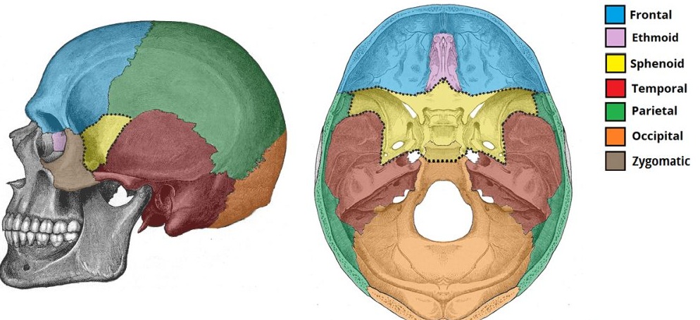
The skull is noticeable from a higher place. The sagittal stitch isolates the left and right parietal bone.
Angles
The front point is a fitting moment and relates to the gathering point of the sagittal and coronal creases. This point is named Brigman. This region is layer-like in the fetal skull and is called front fontanelle for about eighteen months after birth.
The sphenoidal point, flimsy and sharp, is acquired in the space between the frontal bone and the extraordinary arm of the sphenoid. Its inward surface is set apart by a deep furrow, now and again a channel, to the back piece of the center meningeal corridor.
The occipital point is round and relates to the gathering point of sagittal and lambdoidal stitches – a point called lambda; This piece of the skull in the baby is layer and is called back fontanelle.
The mastoid point is small; It is associated with the mastoid piece of the occipital bone and is fleeting. It presents a wide, shallow depression that encases part of the cross-over science on its internal surface. Where this point meets the occipital and worldly mastoid piece is known as the asterion.
Ossification
The parietal bone is solidified into a layer from a solitary place, which shows up on the parietal invulnerability about the eighth seven-day stretch of fetal life.AnglesPoint
The front point is a fitting moment and relates to the gathering point of the sagittal and coronal creases. This point is named Brigman. This region is layer-like in the fetal skull and is called front fontanelle for about eighteen months after birth.
The sphenoidal point, flimsy and sharp, is acquired in the space between the frontal bone and the extraordinary arm of the sphenoid. Its inward surface is set apart by a deep furrow, now and again a channel, to the back piece of the center meningeal corridor.
The occipital point is round and relates to the gathering point of sagittal and lambdoidal stitches – a point called lambda; This piece of the skull in the baby is layer and is called back fontanelle.
The mastoid point is small; It is associated with the mastoid piece of the occipital bone and is fleeting. It presents a wide, shallow depression that encases part of the cross-over science on its internal surface. Where this point meets the occipital and worldly mastoid piece is known as the asterion.
Solidification
The parietal bone is solidified into a layer from a solitary place, which shows up on the parietal invulnerability about the eighth seven-day stretch of fetal life.
Solidification continuously spreads radially from the middle to the edge of the bone. The points bring about the parts that are framed last, and this is in the same place as the fontanels found.
The parietal bone is here and there isolated into two sections, the upper and the lower, by an introduction backstitch.
In different creatures
In barbaric vertebrae, the parietal bones generally structure the back or focal piece of the top of the skull, which lies behind the front bones. In numerous non-well evolved creatures, tetrapods